There is a polypoid , tan colored, ulcerated mass o f the external urethral meatus.
They may then invade nearby structures. Spread of testicular neoplasms 14. Oral malignant melanoma (omm) represents about 1% of all melanomas and approximately 0.5% of all oral malignancies. The evolution of a concept. Oral malignant lesions constituted 14.4% (1,552/10,877).

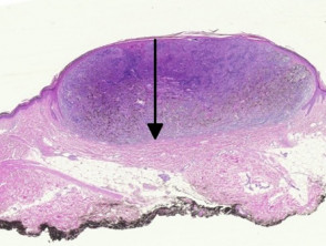
Primary malignant melanoma of the female urethra 207 fig.
(c) a mole that is raised above level of the skin; Omm has been reported in patients aged 20 to 80 years and has a male predilection. Microscopic assessment of breslow thickness is the most accurate prognostic indicator in malignant melanoma. Specimen types include exfoliated cervical cytology (pap tests), urine, body cavity fluids (pleural, pericardial, and peritoneal), cerebrospinal fluid, and fine needle aspirations from any body site, among others (see detail articles section).these are often collected by minimally invasive means. These imaginative details foretold the recent progress made in understanding the pathology of malignant melanoma 18. The appearance of the urethral malignant melanoma at presentation. After 6 years, secondary glaucoma evolved. Though uncommon, soft tissue sarcoma is very dangerous. And (d) a mole that has lost its dark color. The clinical and biologic behavior of desmoplastic melanoma (dm) is a subject of great relevance to those involved in the management of melanoma patients. Benign atypical junctional melanocytic hyperplasia is a descriptive term for a relatively common microscopic alteration. melanoma is the least common form of skin cancer but is the most serious, causing 75% of skin cancer deaths. malignant melanoma develops when the melanocytes no longer respond to normal control mechanisms of cellular growth.
Humoral and cellular autoimmunity play a huge role in the appearance of. The goal of population screening is to detect cancers at their early stages to prevent the associated morbidity and mortality of the late stages. melanoma is a malignant tumor of melanocytes, which are the cells that make the pigment melanin and are derived from the neural crest. malignant melanoma in wales introduction there are two main types of skin cancer: However, melanoma is the most fatal skin malignancy, with an annual mortality approaching 10,000.

Aims to define a retinoinvasive phenotype of uveal melanoma based on an informative case and survey of literature.
Contents functions of the integumentary system the epidermis (thin outer layer of skin) the dermis (thick inner layer of skin) connective tissue and membranes roots, suffixes, and prefixes cancer focus related abbreviations and acronyms further resources functions of the integumentary system. You will find a basic drawing of the layers that make up the skin. Late recurrences of melanoma are well known but uncommon. Pathology report confirmed the diagnosis of malignant choroidal melanoma of the epithelioid type with free optic nerve surgical margin and no signs of vascular invasion. malignant melanoma in wales introduction there are two main types of skin cancer: For comparison's sake, 12,310 new soft tissue sarcomas are estimated to be diagnosed in 2016, while 76,380 new melanomas are expected to be diagnosed, according to the american cancer society (acs). Our goal is to provide a prompt and accurate diagnosis for veterinarians and veterinary ophthalmologists around the world and to advance research for ocular disease in animals. The goal of population screening is to detect cancers at their early stages to prevent the associated morbidity and mortality of the late stages. malignant melanoma is characterized by the development of significant morphological differences and atypical histopathological patterns vs normal cells. Learn about melanoma risk and symptoms and laboratory tests used to evaluate melanoma. Use drawings or photography to note the site(s), size and pigmentation of the lesion(s). malignant melanoma develops when the melanocytes no longer respond to normal control mechanisms of cellular growth. The staging is supplemented visually be line drawings.
For comparison's sake, 12,310 new soft tissue sarcomas are estimated to be diagnosed in 2016, while 76,380 new melanomas are expected to be diagnosed, according to the american cancer society (acs). The efforts to detect melanoma in its early stages went into full gear over 30 years ago. However, a definitive diagnosis of malignant melanoma is a biopsy. Dubow be,ackerman ab, ideas in pathology. The majority of melanomas are black or brown.

How to submit a sample.
Cutaneous foot melanoma is rare, challenging to manage, and not adequately examined in the literature. Light microscopic findings of the surgical specimen. Nodular sclerosis hodgkins lymphoma 12. The evolution of a concept. Our case was presented to draw attention to both the misleading macular appearance of the nm, and its unusual nonhierarchical development. The cell in the skin that contains pigment, is responsible for tanning, and forms melanoma when it becomes malignant. It starts with uncontrolled growth in the cells that make skin pigments. The thin top layer is the epidermis. Contents functions of the integumentary system the epidermis (thin outer layer of skin) the dermis (thick inner layer of skin) connective tissue and membranes roots, suffixes, and prefixes cancer focus related abbreviations and acronyms further resources functions of the integumentary system. This feature was found in 6.2% of otherwise normal intradermal nevi. malignant melanoma is characterized by the development of significant morphological differences and atypical histopathological patterns vs normal cells. 46 patients were identified with a broad range of. malignant melanoma in wales introduction there are two main types of skin cancer:
View Malignant Melanoma Histology Drawing Background. Enzyme markers for myocardial infarction 15. Oral malignant melanoma (omm) represents about 1% of all melanomas and approximately 0.5% of all oral malignancies. malignant melanoma that does not originate in the skin is a very rare disease and is considered one of the most deadly of all human neoplasms. (c) a mole that is raised above level of the skin; With cellular detail similar to that obtained from histology of surgical biopsies).